

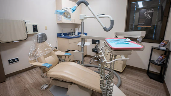
Dental radiographs are essential diagnostic tools that reveal conditions not apparent on clinical examination. They provide detailed visualization of teeth, periodontal tissues and adjacent structures, enabling earlier identification of caries, periodontal disease and other intraoral pathology. Consulting a preventative dentist jacksonville fl helps ensure that imaging is recommended appropriately based on individual risk factors and oral health needs. For patients in Jacksonville, understanding the indications, imaging modalities, recommended intervals and safety protocols supports informed decision-making and helps optimize oral health outcomes. This guide reviews those topics and the factors that influence imaging recommendations.
Guide for Jacksonville Patients on Dental X-rays
Dental radiography is integral to preventive and diagnostic care because it enables timely intervention before conditions progress. In Jacksonville, routine clinical examinations combined with appropriate imaging improve detection and management of oral disease. Patients who understand the role of radiographs can participate in shared decision-making with their dental team about an appropriate imaging schedule.
Necessity of Dental X-rays:
Radiographs detect pathology not visible on visual inspection, including interproximal caries, alveolar bone loss, periapical pathology and developing lesions. Imaging findings inform a comprehensive treatment plan tailored to each patient’s clinical presentation. This proactive diagnostic strategy facilitates earlier treatment and may reduce the need for more extensive procedures later.
Types of Dental X-rays:
Dental radiography comprises several modalities, each selected for a defined diagnostic purpose. The principal categories are outlined below.
- Bitewing X-rays: These are used to detect cavities between teeth and assess the health of the bone surrounding the teeth.
- Periapical X-rays: These provide a view of the entire tooth, from the crown to the root, and are useful for identifying issues below the gum line.
- Panoramic X-rays: These offer a full view of the mouth, including all teeth, jaws, and surrounding structures, making them ideal for treatment planning.
Further research underscores the value of panoramic and single-tooth radiographs for early and accurate diagnosis.
Types of Dental X-Rays for Early Diagnosis
Currently, the most frequent imaging techniques are the orthopantomogram (OPG) and single-tooth radiograph, as well as computer tomography (CT) and cone beam CT mainly for implantology (planning or postoperative control) or trauma indications. Especially early diagnosis and correct classification of a dental trauma, such as dental pulp involvement, prevents from treatment delays or worsening of therapy options and prognosis.
Dental imaging–a basic guide for the radiologist, 2019
What Is a Bitewing X-Ray and When Is It Recommended?
Bitewing radiographs are effective for detecting proximal caries and assessing crestal bone levels in the posterior dentition. Clinicians commonly obtain bitewings during routine examinations, typically at one- to two-year intervals for low-risk adults; frequency is adjusted according to caries risk and clinical findings.
Research supports guideline-based timing for bitewing radiography to improve caries detection.
Dental X-Ray Frequency: Guidelines for Patients
ObjectivesTo identify guidelines on when and how frequently bitewing radiographs should be used in dentistry for the diagnosis of caries, and to provide an objective appraisal of their quality. Publications were included if they made recommendations on the issue of when and how frequently radiographs should be used in any dentally-related specialty pertaining to the diagnosis of caries; and/or if they were aimed at the individual practitioner (any health professional working within dentistry) and/or patients.
Guidelines on the timing and frequency of bitewing radiography: a systematic review, H Devlin, 2017
How Do Panoramic and Digital Dental Radiography Enhance Diagnosis?
Panoramic radiography produces a single image of the maxillofacial region, useful for assessing dentition, jaw relationships and occult pathology. Digital intraoral sensors deliver immediate images at a lower radiation dose than film, enabling faster diagnosis and more efficient treatment planning.
Frequency of Dental X-rays:
Recommended imaging intervals depend on individual risk assessment, dental history and clinical indicators. Generally, adults at low risk may require radiographs every one to two years; children and higher-risk patients often need more frequent imaging to monitor development and disease progression. The dentist will determine the optimal schedule for each patient.
| Patient Group | Recommended Frequency | Reason |
|---|---|---|
| Adults | Every 1-2 years | Routine monitoring of oral health |
| Children | Every 6-12 months | Monitoring growth and development of teeth |
| High-risk patients | Every 6-12 months | Increased likelihood of dental issues |
At Ramos Dental Care PA, we emphasize individualized care and set each patient’s radiographic schedule to reflect their specific oral health status and risk profile.
Safety Measures:
Contemporary dental radiography is designed to minimize patient dose. Digital sensors, optimized exposure settings and protective barriers such as lead aprons and thyroid collars are standard practice. Together, these measures ensure diagnostic imaging is performed with minimal risk.
Further research underscores the importance of comprehensive radiation protection measures in dental radiography.
Understanding Dental X-Ray Safety & Radiation Protection
X-rays cause ionization of atoms, which is one factor contributing to the harmful effects of X-radiation in living organisms. Humans are exposed to radiation from various sources, and medical exposure may not be the primary contributor. Radiation protection encompasses measures aimed at preventing exposure and contamination of the natural environment, and when prevention is not possible, at minimizing effects to the lowest achievable level while considering economic, sociological and health factors.
Safety Precautions for Dental Patient and Dental Staff Using X-Rays, 2020
Factors Influencing Recommendations:
Recommendations for radiographic frequency reflect multiple factors: previous caries experience, periodontal status, developmental stage, systemic health and clinical findings. Patients with recurrent caries or active periodontal disease typically require closer radiographic surveillance than those with stable, low-risk dentitions.
What Are the Different Types of Dental X-Rays Used in Preventative Care?
Preventive protocols employ intraoral bitewings, periapical radiographs and panoramic imaging as indicated to monitor caries, root and periodontal health and tooth development. Each modality contributes specific diagnostic information that supports targeted preventive treatment.
How Safe Is Digital Dental Radiography for Patients in Jacksonville?
Digital radiography is regarded as a safe imaging option because it substantially reduces radiation dose compared with conventional film systems. When combined with standard protective practices, digital methods allow clinicians to obtain required diagnostic information while minimizing patient exposure.
What Are the Radiation Exposure Levels Compared to Traditional Methods?
Digital intraoral radiography typically delivers markedly lower effective doses than film-based techniques; studies report dose reductions ranging approximately from 50% to 90%, depending on the system and technique. This reduction is particularly relevant for children and other radiation-sensitive groups.
One study quantified this reduction, demonstrating a significant decrease in radiation exposure with digital systems.
Digital Radiography: 55% Dose Reduction Compared to Film
digital systems reported a dose reduction of 55% on average compared with film. This is a dose reduction state and photostimulable phosphor systems for digital intra-oral radiography.
The dynamic range of digital radiographic systems: dose reduction or risk of overexposure?, WER Berkhout, 2004
This significant reduction in radiation exposure is further supported by studies comparing digital and film-based systems.
Digital vs. Film Dental X-Rays: Patient Dose Reduction
Despite a slight increase of radiographs taken with CCD systems, there is a significant dosage reduction. Corresponding to the decrease in exposure time, the patient dose for SPP systems is reduced to one half compared to film.
Intraoral radiology in general dental practices–a comparison of digital and film-based X-ray systems with regard to radiation protection and dose reduction, 2014
What Safety Guidelines Does Ramos Dental Care Follow?
Ramos Dental Care PA adheres to established radiation safety standards. Our protocols include appropriate use of digital sensors, routine equipment maintenance, application of protective shielding and ongoing staff training in exposure optimization and patient safety.
How Often Should Different Patient Groups Receive Dental X-Rays?
Imaging frequency should be customized by the treating clinician. Age, caries risk, periodontal condition and previous treatment history inform the recommended interval for radiographs for each patient group.
What Are the Frequency Recommendations for Adults Based on Oral Health Risk?
For adults, typical recommendations range from one to two years for routine bitewing imaging. Adults with elevated caries risk or active periodontal disease may require radiographs every six to twelve months to monitor treatment response and disease progression.
When Should Children and Pediatric Patients Have Dental X-Rays?
Children generally require more frequent radiographic assessment because of tooth eruption and higher caries susceptibility. Clinicians commonly schedule imaging every six to twelve months as clinically indicated to track development and identify emerging problems early.
What Role Does the Comprehensive Oral Exam Play in Scheduling Dental X-Rays?
A comprehensive oral examination provides the clinical context for radiographic decisions. Findings from the exam determine whether imaging is indicated and which modality will yield the most relevant diagnostic information.
How Does the Oral Exam Inform Personalized X-Ray Frequency?
The clinical assessment guides a personalized radiographic plan by integrating the patient’s dental history, current pathology and risk factors. This ensures imaging is performed only when it will influence diagnosis or treatment.
How Are Diagnostic Imaging and Preventative Care Integrated?
Diagnostic imaging supports preventive care by revealing early lesions and anatomic conditions that clinical examination alone may miss. Timely identification of such issues enables conservative interventions and improves long-term oral health outcomes.
What Are Common Patient Questions About Dental X-Ray Frequency and Safety?
Patients frequently ask about the necessity and safety of radiographs. Clear, evidence-based explanations help patients understand the clinical value of imaging and the measures taken to protect them.
Are Dental X-Rays Safe for Pregnant Women and Children?
When clinically necessary, dental radiographs may be performed for pregnant patients and children with appropriate precautions. Use of digital sensors, lead aprons and thyroid collars reduces exposure. Pregnant patients should inform their clinician so that imaging can be deferred or modified when appropriate.
How Much Radiation Do Dental X-Rays Emit Compared to Everyday Exposure?
The effective dose from a single dental radiograph is low compared with natural background radiation. For perspective, many intraoral exposures approximate only a few days’ worth of ambient background dose, which supports their favorable risk-benefit profile for diagnostic purposes.
How Can Jacksonville Patients Prepare for Their Dental X-Ray Appointments?
Preparation is minimal. Patients should inform the dental team of pregnancy, recent imaging or relevant medical history. Maintaining routine oral hygiene prior to the visit facilitates efficient imaging and assessment.
What Should Patients Expect During a Dental X-Ray Procedure?
During intraoral radiography, the patient will be seated and may be asked to bite on a sensor or film holder while the operator positions the X-ray head. The exposure itself is brief and the overall procedure typically requires only a few minutes.
How Does Ramos Dental Care Ensure Patient Comfort and Safety?
At Ramos Dental Care PA, our team follows established protocols to maintain patient comfort and minimise exposure. We employ modern imaging equipment, protective shielding and patient‑centred communication to ensure a safe and supportive experience.
To ensure you are getting the best preventative care, it is important to understand all of your options. Preventative care is a key part of maintaining good oral health.
Frequently Asked Questions
What Are the Long-Term Effects of Dental X-Ray Exposure?
Dental X-rays involve exposure to low levels of ionizing radiation; when proper safety protocols are followed, long-term effects are minimal. The risk of radiation-related health issues is extremely low, particularly with modern digital imaging that reduces exposure. Regular dental check-ups and appropriately timed radiographs are important for maintaining oral health, and the diagnostic benefits typically outweigh the risks. Discuss any concerns with your dentist for personalized guidance.
How Do I Know If I Need Dental X-Rays?
The need for dental radiographs is determined by your dentist based on oral health history, current symptoms and risk factors. During routine examinations, the clinician will assess whether imaging is required to diagnose potential problems. Age, previous dental issues and overall health influence this decision. Communicate specific concerns or symptoms to receive tailored advice.
Can Dental X-Rays Detect Issues Beyond Cavities?
Yes. Dental X-rays detect cavities and a range of other conditions, including periodontal disease, bone loss, abscesses, tumors and the position of teeth such as impacted third molars. This comprehensive diagnostic information allows dentists to develop effective treatment plans and monitor oral health. Regular imaging supports early detection and intervention to prevent more serious complications.
What Should I Do If I Have a History of Radiation Sensitivity?
If you have a history of radiation sensitivity or related health concerns, inform your dentist before undergoing X-rays. The clinician can evaluate your situation, consider alternative imaging methods or adjust the frequency of radiographs, and ensure all appropriate safety measures are in place. Open communication with your dental team is essential to protect your health.
How Do Dental X-Rays Contribute to Preventive Care?
Dental X-rays are central to preventive care because they reveal early lesions and structural changes that clinical examination may miss. Early detection enables conservative treatment, helps maintain oral health and reduces the likelihood of more extensive and costly procedures in the future.
What Are the Guidelines for X-Ray Frequency in Special Populations?
Special populations have specific guidelines for radiographic frequency. Pregnant women should receive X-rays only when clinically necessary and with precautions to minimize exposure. Children, because of developing dentition and higher caries susceptibility, may require more frequent imaging, typically every six to twelve months. Dentists will tailor recommendations to individual health needs and risk factors to ensure safe care.
What Are the Risks of Dental X-Rays?
Dental radiographs involve a small ionizing radiation dose. With modern digital systems and appropriate protective measures, the associated risk is minimal. Clinicians balance the low exposure against the diagnostic benefits, particularly when monitoring active disease or planning treatment. Discuss individual concerns with your dentist to ensure appropriate care.
How Can Patients Reduce Their Need for Frequent X-Rays?
Maintaining effective oral hygiene, attending scheduled examinations and addressing carious lesions or periodontal disease promptly can reduce the need for additional imaging. Adherence to preventive recommendations lowers risk and supports less frequent radiographic surveillance when clinically appropriate.
What Should Patients Do If They Have Concerns About X-Ray Frequency?
Patients should raise questions with their dentist and request an explanation of the clinical indication for each radiograph. Dentists will provide personalized recommendations based on history, current findings and risk factors to ensure imaging is justified.
Are There Alternatives to Dental X-Rays?
Certain adjunctive methods, such as transillumination or laser fluorescence, can support caries detection in selected cases. However, these alternatives do not always provide the comprehensive anatomic information that radiographs deliver. The dentist will advise on the most appropriate diagnostic approach for the clinical situation.
How Do Dental X-Rays Impact Treatment Planning?
Radiographs supply critical information about tooth roots, periapical tissues, bone levels and hidden caries, enabling clinicians to formulate accurate, targeted treatment plans. Reliable imaging contributes to timely, effective interventions and improved clinical outcomes.
What Should Patients Know About X-Ray Technology Advancements?
Advances in digital radiography have substantially improved safety and workflow. Digital systems reduce radiation dose compared with film, provide immediate images for rapid assessment and facilitate secure electronic storage and interdisciplinary collaboration. These developments enhance the quality and efficiency of dental care.
Conclusion
Understanding when and why dental radiographs are used helps patients make informed choices about their oral care. Radiographic imaging supports early detection, personalized treatment planning and safe, effective preventive management. Consult your dentist to establish an imaging schedule that reflects your clinical needs and optimizes long-term oral health.


